Infrared-light coagulation: Mode of functioning
Infrared-Light Coagulation
is a very reliable, rapidly effective and simple-to-use method for haemostasis.
The selected probe is placed on the tissue that needs coagulating and gentle pressure applied. The user then triggers an intensive infrared-light impulse which penetrates the tissue, is absorbed there and causes a coagulation of up to 8 mm depth within a few seconds by means of heat conversion.
Application of the Infrared-Light Coagulator
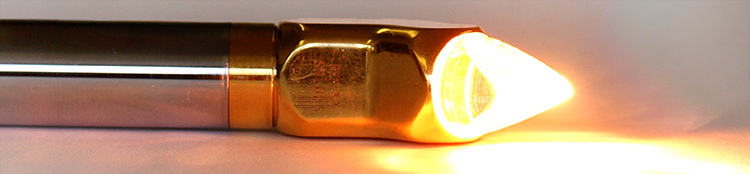
 The tip of the probe is gently pressed onto the bloddy tissue surface (contact coagulation). This already leads to a certain degree of haemostasis which prevents further blood from flowing. After that, a timed intensive light impulse is triggered.
The rays penetrate several millimeters into the bleeding tissue at the speed of light, are absorbed there and converted into heat.
Depending on the duration of application and the tissue structure, sufficient temperature is reached after a few seconds achieving the following effects on the tissue:
| Effect | Temperature | Duration of Impulse | Coagulation Depth |
|---|---|---|---|
| Denaturalisation | at approx. 50-60°C | approx. 0,5-2,0 sec | approx. 1-2 mm |
| Evaporation of Cell Water | at approx. 100°C | approx. 2,0-3,5 sec | approx. 2-3 mm |
| Collagen (Glue) | at approx. 170°C | approx. 3,5-5,0 sec | approx. 3-5 mm |
| Carbonisation | at approx. 300°C | over approx. 5,0 sec | max. approx. 5-7 mm |
 The diameter of the coagulated are corresponds approximately to the diameter of the contact area. The coagulation depth corresponds approximately in millimeters to the coagulation period in seconds.
Advantages of the Infrared-Light Coagulation compared to other methods
- Both the area and depth affected by the infrared-light coagulation are confined by a controlled light emission. Adjoining or subjacent tissue is therefore not affected and there is no danger of perforation.
- The effect which is achieved in seconds guarantees reliable haemostasis. This results in less consumption of blood conserves, shorter operation times and time saving in emergencies.
- Through the combination of light and tissue contact the infrared-coagulator performs reliably when dealing with diffuse, extensive bleeding or where exact origin of bleeding is unknown. Haemostasis is possible at any time, particularly without preparatory measures such as vacuum-pumping or prior blotting of the area – even under a pool of blood.
The use of ‘infrared-light’
prevents both side-effects and additional costs:
- No danger of patients suffering irritation or burns from electrical current
- No additional running costs such as expensive fibrin glue for wound closures or technical gases for cooling purposes
- No elaborate safety measures needed
- No smoke formation – no visual obstruction
- Pain-free coagulation when taking biopsies

Anwendung EKA GmbH Infrarot-Lichtkoagulator Beispielvideo 01

Anwendung EKA GmbH Infrarot-Lichtkoagulator Beispielvideo 02

Anwendung EKA GmbH Infrarot-Lichtkoagulator Beispielvideo 03

Anwendung EKA GmbH Infrarot-Lichtkoagulator Beispielvideo 04

Anwendung EKA GmbH Infrarot-Lichtkoagulator Beispielvideo 04a